Whole-body MRI could detect minimal residual disease, predicting multiple myeloma relapse
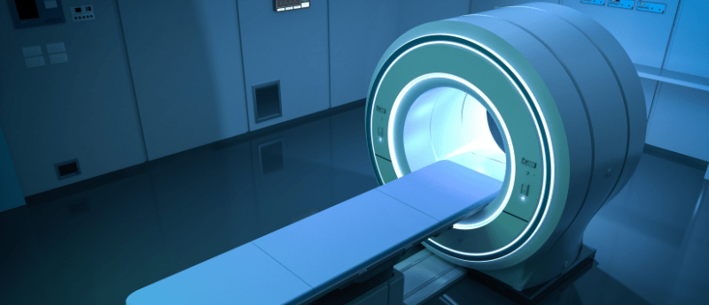
Whole-body MRI scans have been shown to detect residual multiple myeloma after treatment, even when traditional blood and bone marrow tests show no signs of cancer.
For the first time, whole-body MRI (WB-MRI) has been used to predict treatment outcomes in patients with multiple myeloma (MM). When used with a standardized set of criteria to interpret and report WB-MRI, researchers from The Royal Marsden NHS Foundation Trust and The Institute of Cancer Research (both London, UK) were able to detect residual disease after treatment, potentially enabling better predictions of potential relapse.
Currently, treatments for MM focus on controlling symptoms extending remission and improving quality of life outcomes; however, it cannot be cured. Although patients are monitored post-treatment with blood tests, bone marrow biopsies, CT scans and X-rays, residual disease can be missed. By the time any secondary cancer has been found, it is often too late.
A new tool for predicting relapse
In this study, the researchers investigated whether WB-MRI and the Myeloma Response Assessment and Diagnosis System (MY-RADS) could be used to evaluate residual MM in patients, following their treatment.
The team followed 70 patients undergoing a stem cell transplant, conducting WB-MRI before and after. WB-MRI indicated residual disease in approximately one third of the patients, who went on to have a median progression–free survival of just 24 months. Overall survival was also shorter. Comparatively, the remaining patients whose WB-MRI didn’t indicate residual disease had a median progression–free survival of 42 months.
Towards smarter, kinder diagnostics
Christina Messiou (The Royal Marsden NHS Foundation Trust and The Institute of Cancer Research) study chief investigator, explained: “This study shows that WB-MRI gives us valuable information about how well the myeloma has responded to treatment that other tests may miss. It’s exciting that we now have a standardized, non–invasive imaging method that can be used across cancer centres. WB-MRI doesn’t involve radiation or intravenous injections, which is important for patients who may require lifelong monitoring. This is an important step towards smarter and kinder precision diagnostics for patients with cancer.”
Martin Kaiser (The Institute of Cancer Research and The Royal Marsden NHS Foundation Trust) commented: “Access to this gold-standard precision imaging with WB-MRI has revolutionized care for myeloma patients. As the treatment options for myeloma increase and factors such as disease distribution across the body are increasingly understood as important to treatment response, the relevance of the whole body-MRI for personalizing treatment will only increase over time.”
The team hopes that as imaging techniques advance, they could become part of standard care and allow more patients to receive personalized treatments for their cancer.