New imaging software could improve breast cancer diagnosis
Newly developed software could provide a faster breast cancer diagnosis with 90% accuracy and without the need for specialist pathologists. Investigators suggest that this development could be particularly valuable in resource-poor settings and developing countries. The work was published recently in the journal Breast Cancer Research.
“To evaluate fresh breast tissue at the point of care could change the current practice of pathology,” reported study author Rebecca Richards-Kortum (Rice University, TX, USA). “We have developed a faster means to classify benign and malignant human breast tissues using fresh samples and thereby removing the need for time consuming tissue preparation.”
Breast cancer diagnosis is currently a complex procedure. Tissue must firstly be obtained by surgical excision or core needle biopsy. Following this, the sample is then meticulously prepared before undergoing a lengthy evaluation and diagnosis.
The research team, by contrast, analyzed intact breast tissue samples using high-speed optical microscopy. This automated method foregoes the need for specialists to undertake intricate sample preparation and assessment.
Richards-Kortum continued: “We performed our analysis without tissue fixation, cutting and staining and achieved comparable classification with current methods. This cuts out the tissue preparation process and allows for rapid diagnosis. It is also reliant on measurable criteria, which could reduce subjectivity in the evaluation of breast histology.”
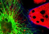
The novel program scrutinizes images of breast tissue samples, from a confocal fluorescence microscope, according to criteria used in tissue analysis. The results are then entered into a classification tree designed by the research team to determine whether the tissue sample is malignant or benign.
There are, however, limitations to the software. Typically, optical microscopy is not used in patient care due to its high cost and certain diagnostic parameters are subject to user error.
Jessica Dobbs (Rice University) concluded: “We are excited about the possibility to use these imaging techniques to improve access to histologic diagnosis in developing regions that lack the human resources and equipment necessary to perform standard histologic assessment.”
Source: BioMed Central press release
fsgadmin
