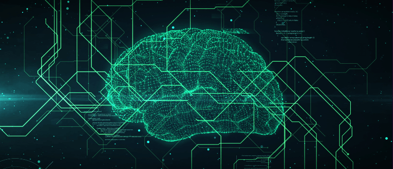
How can AI help provide real-time insights into glioblastoma disease progression?
 Dr Radka Stoyanova is Professor and Director of Imaging and Biomarkers Research at the Sylvester Comprehensive Cancer Center, Miller School of Medicine, University of Miami (FL, USA). She is currently working on an innovative approach to trace glioblastoma tumors in large MRI datasets using machine learning. In this interview, Stoyanova speaks with Senior Editor Jade Parker about how this trailblazing research could be applied in clinics and key opportunities for women in brain tumor research.
What advantages does MRI-guided radiation therapy offer in terms of real-time insights into glioblastoma disease progression?
Acquiring MRI during radiation treatment (RT) provides a unique glimpse into the cancer’s evolution. The ability to gather information on glioblastoma response earlier than standard imaging (at 3 months post-RT) is crucial for timely clinical management adjustments, especially in rapidly growing tumors such as glioblastoma. The contemporary MRI-guided RT platforms acquire daily MRI scans that provide an accurate anatomical characterization of the changes both in the tumor and the surrounding brain tissues. An exciting potential benefit is the analysis of serial MRIs to monitor treatment response or predict outcomes. My research group focuses on developing approaches for optimal extraction of information from large imaging datasets and utilizing longitudinal MRI data to enhance data mining. In addition to detecting the tumor volume changes, a series of quantitative imaging features can be extracted from the underlying image and using this process, referred to as radiomics, we can build models to predict the response of a patient to a particular treatment. In addition to the tumor, analyzing the radiomics features derived from the “normal-appearing” brain tissues may provide further important information for the changes in the tumor environment that may be a crucial contributing factor to the overall response and patient outcomes.
Can you elaborate on how artificial learning is applied to determine whether the tumor is shrinking or growing during the treatment process?
Our preliminary data from a limited patient set suggests differential patterns of tumor volume dynamics during RT related to the patient’s response/resistance to treatment. Of note, there are 30 three-dimensional datasets acquired during RT of glioblastoma. The manual contouring of the tumor on multiple slices is time consuming. On average, 30−60 minutes are needed to contour a single exam, which results in 15−30 hours per patient. Moreover, some of the cases are quite challenging and require more input and hence the valuable time of an expert radiation oncologist specialized in brain tumors to help in contouring. While software packages for automatic segmentation of glioblastoma exist, they were developed on data acquired on a stronger magnet and from multiple sequences that are not available on the MRI-guided RT platform. The developed deep learning network will allow the rapid collection of data that will further strengthen our initial observations for differential patterns of tumor volume dynamics.
 Uncovering cancer’s secrets: could liquid biopsies and artificial intelligence hold the answers?
Future Oncology recently published this Editorial exploring the potential of AI and liquid biopsies in achieving early cancer detection.
How would you like to see this research applied in clinic?
The developed network can segment a single dataset in less than 3 minutes thereby enabling real-time monitoring of the changes in tumor size and anatomy. We anticipate that a rapid assessment of treatment response will be possible based on the derived tumor volume growth/shrinkage patterns, together with the developed radiomics model. We anticipate that this dynamic system will be implemented on the MRI-guided RT platform and will be used for flagging patients with favorable/unfavorable response for corresponding deintensification or intensification of the treatment.
What motivated you to pursue a career in oncology and how have you overcome challenges along the way?
My fascination with medical imaging as a window to complex structures and disease phenotypes has been a driving force in my career. Throughout my scientific journey, I’ve focused on understanding cancer-related data for more accurate retrieval of information. Working closely with clinical colleagues, I’ve learned to navigate challenges by being flexible, understanding clinical needs, and effectively communicating the applicability of our tools in clinical settings.
Are there specific areas within brain tumor research where you see opportunities for more women to make significant contributions?
Personalized and targeted treatment strategy development, particularly through non-invasive biomarkers from imaging, presents significant opportunities for women in brain tumor research. As far as brain tumors are concerned, non-invasive biomarkers from imaging and other technologies are of paramount importance as they eliminate the need for surgical procedures to obtain tissue samples directly from the tumor. One of the most promising areas of brain research is the so-called “liquid” biopsies. Liquid biopsies refer to a non-invasive diagnostic technique that involves analyzing various biomarkers, such as circulating tumor cells (CTCs), cell-free nucleic acids (cfDNA), and extracellular vesicles present in blood and/or other body fluids, including cerebrospinal fluid (CSF) for brain tumors. In the context of cancer, liquid biopsies are used to detect and analyze genetic mutations, alterations, or other molecular changes associated with tumors. Liquid biopsies, together with imaging offer the advantage of being able to capture the phenotypic and genetic heterogeneity of tumors and monitor the disease’s progression or response to treatment over time.
How important has mentorship been in your career? Have you had any notable mentors or role models who influenced your journey?
Mentorship has played a crucial role in my career. I was introduced to the field of imaging by two women who later became titans in the field: June Taylor and Sarah Nelson, formerly from St. Jude Children’s Hospital (TN, USA) and University of California, San Francisco (CA, USA). Both of them were mavericks in the field of neuro-quantitative imaging analysis and I am eternally grateful for their efforts and struggles that paved the way for the women that followed. As my career progressed, mentorship became a focus, and I actively contribute to mentoring junior faculty, students, and colleagues, fostering scientific curiosity, ethical standards, and career development.
The opinions expressed in this interview are those of the author and do not necessarily reflect the views of Oncology Central, Neuro Central or Taylor & Francis Group.