Developing a ‘Google Earth’ for tumors

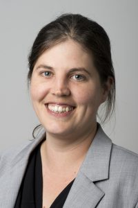
In an exclusive interview, Josephine Bunch of the National Physical Laboratory (NPL; London, UK), speaks to Oncology Central about her project which will utilize mass spectrometry imaging to create a Google Earth–like tumor map. The ground-breaking project has been selected by Cancer Research UK to receive £16 million over the next 5 years as part of its Grand Challenge awards; set up to revolutionize the prevention, diagnosis and treatment of cancer and help scientists tackle unanswered questions in cancer research.
Can you tell our readers about your career & what led you to where you are today?
I completed a PhD in mass spectrometry imaging at Sheffield Hallam University (UK), sponsored by Pfizer Global R&D, before moving to the Department of Chemistry at the University of Sheffield for my postdoctoral appointments. During this time, I was awarded an independent Enterprise Fellowship to support commercialization of mass spectrometry imaging activities. I then moved to the University of Birmingham (UK), where I led a research group in mass spectrometry imaging and was a Lecturer in Chemistry and Imaging in the School of Chemistry and Physical Sciences of Imaging in the Biomedical Sciences (PSIBS) Doctoral Training Centre. I joined the National Centre of Excellence in Mass Spectrometry Imaging at the National Physical Laboratory in 2013, to lead research in MALDI and ambient mass spectrometry imaging.
You are the Principal Scientist and Co-Director of the National Centre of Excellence in Mass Spectrometry Imaging. Could you tell us a bit about the center and the role mass spectrometry plays in cancer research?
NiCE-MSI at NPL is one of the world’s leading mass spectrometry imaging (MSI) centers with a focus on advancing measurement capabilities, establishing metrology for reliability and standardization, supporting UK industry, and training the next generation of scientists and engineers.
Mass spectrometry imaging (MSI) allows molecular chemistry to be visualized in 2D and 3D, from the nano- to the macro-scale, in ambient conditions as well as in real time, and has been utilized to study the distribution of proteins, lipids and drugs in a diverse range of tissues and cells. As such, MSI offers unique opportunities in cancer treatment for measuring disease markers, drug distribution and importantly the molecular changes occurring within tumors throughout treatment. These attributes highlight the excellent potential for MSI to inform treatment decisions and monitor treatment responses to help the development and delivery of therapies that are effective for particular groups of patient.
Josephine recently won the Cancer Research UK Grand Challenge competition, designed to answer the biggest questions in cancer research. Can you tell us what winning this competition meant to you and your team?
This is the most exciting project I have ever been involved with. The scale of the funding offered by Cancer Research UK has allowed us to put together an extremely ambitious program of research, which spans new and exciting work in both physical and life sciences. Across the consortium, we have a strong track record of delivering successful research through a number of smaller multidisciplinary projects and we’re very excited about this new project. We will measure things which have never been measured, using the most advanced instruments in the world and by doing this we will generate new insights into tumor metabolism and cancer progression. We hope that this will lead to new ways of diagnosing and treating cancer.
Your Grand Challenge project will focus on developing a ‘Google Earth’ for tumors to improve cancer diagnosis and treatment.
a. Could you give us a brief overview of this project?
The 5-year project brings together physical scientists who have innovated next-generation mass spectrometry imaging techniques and instruments with international experts in cancer biology to address Cancer Research UK’s Grand Challenge of ‘mapping tumors at the molecular level’. By developing a standardized way to fully map tumors in unprecedented detail – from the whole tumor to the individual molecules in cells – we’re aiming to deepen understanding of the interplay of genes, proteins, metabolites and the role of the immune system in cancer development and growth.
b. What methods will you use to map tumors?
We will utilize a range of MSI techniques (MALDI, desorption electrospray ionisation mass spectrometry (DESI MS), secondary ion mass spectrometry (SIMS), inductively coupled plasma mass spectrometry (ICP-MS) and rapid evaporative ionization mass spectrometry (REIMS)), as well as magnetic resonance spectroscopy and imaging. This will allow us to build a comprehensive, spatially resolved picture of the metabolism of solid tumors from subcellular to anatomical levels using in-vivo, ex-vivo and in-vitro analyses.
c. What do you hope the impact of this research will be?
The knowledge provided will help define new therapeutic strategies, identify novel targets for drug development and provide more accurate methods for diagnosis and monitoring treatment. Ultimately, it could help more individuals survive cancer for longer.
d. What are your long-terms goals for the application of this model?
It’s very important to us that this project delivers methods that are reliable and can be utilized by other laboratories. We are going to create a searchable database that will be freely available to researchers around the world, to help create a standardized way for other scientists and doctors to use these novel mass spectrometry imaging techniques in their work and in the clinic.
Over the next 10 years what changes would you like to see in the field of cancer research?
Although reproducibility in research has problems across all disciplines, as highlighted in the recent POSTnote on research integrity [1], the implications of this in healthcare research are particularly troubling. Recently, the first results of the Reproducibility Project, which attempts to independently replicate the results of landmark cancer studies, suggested that only two of the first five studies attempted were reproducible [2-3].
Over the next 10 years, I would like to see this change. I want to see more focus on the adoption of working practice and reporting guidelines that ensure the work we do is reproducible. When utilizing complex techniques on complex systems, as is especially the case in cancer research, it is vital we think about how to make the experiments we carry out and the data we generate as useful as possible, and ultimately, bring the most benefit to patients. As the UK’s National Measurement Institute, we at NPL can provide the standardized methods, tools and protocols needed to achieve this.
Do you have any closing message for our readers?
In awarding our Grand Challenge project, Cancer Research UK is recognizing the importance of fostering new multidisciplinary collaborations to tackle society’s biggest challenges. NPL fulfils a key role in this multidisciplinary approach, ensuring we’re making quality measurements that lead to meaningful, reproducible results that can make a real difference to people’s lives.
References:
[1] Parliamentary Office of Science and Technology: Research Integrity http://researchbriefings.parliament.uk/ResearchBriefing/Summary/POST-PN-0544?utm_source=website&utm_campaign=PN544
[2] NosekBA, Errington TM; Reproducibility in cancer biology: Making sense of replications eLife.
doi: 10.7554/eLife.23383 (2017)
[3] Baker M, Dolgin E; Cancer reproducibility project releases first results. Nature. doi: 10.1038/541269a (2017).
Biography
Dr Josephine Bunch is Co-Director of the National Centre of Excellence in Mass Spectrometry Imaging (NiCE-MSI) at the National Physical Laboratory and Associate Professor in the School of Pharmacy, University of Nottingham (UK). Josephine has expertise in a range of mass spectrometry imaging techniques and leads research in matrix-assisted laser desorption/ionization (MALDI) and ambient mass spectrometry imaging at NPL.
