Novel imaging-based method could help optimize breast cancer therapy
A novel imaging-based method developed to aid in determining suitability of HER2-targeted therapies in breast cancer had been demonstrated to be as accurate as the current standard-of-care in a new study. The findings, published in the journal Theranostics, may lead to diminished use of invasive tissue samples and could help optimize breast cancer therapy.
The study, carried out by investigators at Uppsala University (Sweden), aimed to develop a noninvasive method for the determination of HER2 levels within metastatic breast tumors – elevated levels of which are currently identified based on examination of tissue samples obtained by surgery or needle biopsies.
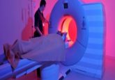
The new method utilizes a whole-body PET/CT imaging technique using the novel tracer ABY-025 Affibody, which is labeled with short-lived radioactive isotope gallium-68. Sixteen women with metastatic breast cancer received the test, the results of which were then compared with their HER2 measurements from invasive biopsies.
Following analysis, it was shown that HER2-expression levels in the metastases were accurately measured with the PET/CT method. In addition, HER2 expression within metastases was frequently found to be different from the primary tumor, leading to therapy changes for several patients.
The authors postulated that this new method may have the potential to substitute invasive tissue sampling in the near future. They now plan to confirm their results in a larger group of patients from multiple hospitals with an aim of making the new method more widely available to patients.
Source: Uppsala University press release