Glowing for gold: early glioma detection using fluorescent probes
Scientists at Hokkaido University have pioneered a revolutionary non-invasive method to assess the malignancy grade of glioma tumor cells, potentially transforming early cancer detection.
New research, led by Yasuchika Hasegawa and Shinya Tanaka of the Institute for Chemical Reaction Design and Discovery at Hokkaido University (Sapporo, Japan), have developed a non-invasive probing model for assessing the malignancy grade of glioma tumor cells. The approach uses a water-soluble, luminescent europium Eu(III) complex, offering a promising avenue for early cancer detection.
Glial cells are connective tissue supporting the neurons within the brain and spinal cord. Accounting for over a quarter of brain cancers, glioma affects healthy glial cells. Glioma brain tumors vary across the spectrum of low-grade (slow-growing) and to high-grade (faster-growing) with high-grade tumors generally having a poorer prognosis.
Early detection and diagnosis are key to increase the chances of successful cancer treatment. However, current methods of evaluating brain tumor malignancy are often invasive with high-risk complications.
Bioimaging technologies based on luminescent molecules have emerged as a tool to localize and distinguish active cancer cells. Among the luminescent molecules, transition metal complexes hold a particular advantage in bioimaging and cancer diagnosis due to their extended phosphorescence lifetimes. Hasegawa and Tanaka focused their research on harnessing the unique properties of a structure-changeable Eu(III) complex to detect the activities of early human glioma tumor cells.
This case report describes the case of a patient diagnosed with MGMT-methylated glioma who received cilengitide in the CENTRIC clinical trial.
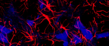
The study employed three different model cells which mimic varying grades of malignancy. By measuring the lifetime of the Eu(III) complex’s characteristic red light emission, researchers observed significant differences in the behavior of more malignant cells within the initial three hours.
Hasegawa emphasized, “visualization of cancer cells using luminescent complexes has previously been reported, but our hypothesis was that the photophysical signals sent by such complexes in cancer cells might reflect internal information from the cancer cells.”
To achieve the characteristic red-light emission, the research team first modified the Eu(III) complex to ensure water solubility and stability among the amino acids in the cell culture mediums. In the cell medium, it initially forms an aggregate with other Eu(III) molecules. Upon interaction with model tumor cells, the aggregate breaks into single Eu(III) molecules, which are rapidly taken up by the cancer cells. This process promotes structural changes resulting in the red-light fluorescence.
This research represents a significant leap forward in the field of cancer detection and diagnosis. By offering a non-invasive method to assess the malignancy grade of glioma tumor cells, the study opens new possibilities for early intervention and improved treatment strategies. Despite this, further research is needed to evaluate the probing ability of Eu(III) in vivo.