Breakaway cancerous cells successfully mapped and tracked with metal detection
Researchers have successfully utilized a novel metal detection technique for mapping and tracking breakaway cancerous cells. The findings, published recently in Convergent Science Physical Oncology, may help researchers develop more precise treatment plans for individual patients.
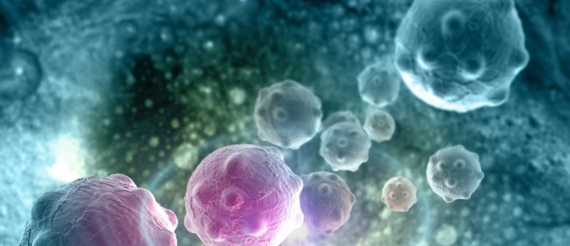
The team from USC Michelson Center (CA, USA) achieved the above by imaging metal-tagged antibodies on biopsies from a patient with metastatic prostate cancer; in order to create highly detailed, digital copies of cancer cells that can travel through the body.
The metal tags enabled the team to identify and characterize the cancer cells in a blood sample after it was placed on a slide.
“We are trying to understand how cancer actually moves from the initial location to other places in the body and can settle there,” lead author Peter Kuhn from USC Michelson Center, explained.
Up until now, researchers have relied on fluorescence microscopy, which though useful, is limited in the number of colors available in a single experiment.
The new approach of using metal-tagged antibodies and a laser ablation system, coupled with a mass spectrometer, allowed the scientists to view circulating and disseminated tumor cells at a molecular level in a way not previously possible. It provided the team with the ability to track 35 different metal labels simultaneously; overall, producing 35 distinct views of the cancer cell’s biology.
The team hopes that by creating such highly detailed copies of tumors they will be able develop more precise treatment plans for individual patients.
“This is really just the beginning,” Kuhn commented. “You’ll see hundreds of studies now using this technique.”
Source: www.eurekalert.org/pub_releases/2018-02/uosc-smt022718.php