Vote for your winner of the Capturing Cancer: Oncology Central Photography Competition
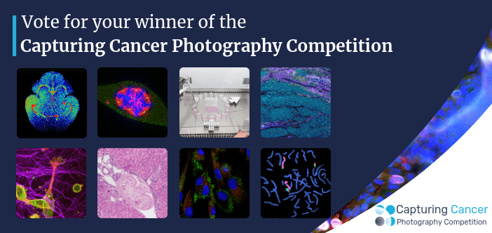
Our panel of expert judges have chosen their top photos from all of the entries and the public have casted their votes, find all the finalists for our competition below.
The winners will receive:
- US $100 Amazon voucher
- A whopping 35% off Open Access when they submit a paper to the journal Future Oncology or 50% off Open Access when they submit a paper to one of our Oncology Management journals
- The opportunity to showcase their work to the global oncology community through an exclusive interview, with their image being showcased on our homepage image slider
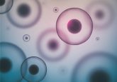
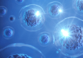
Mesenchymal Stem Cells
Photographer: Volha Vshyukova
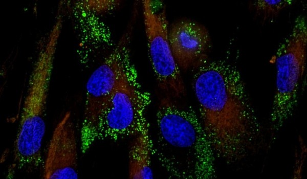
Baculovirus transduction of human mesenchymal stem cells
In Extracellular space no one can hear you scream
Photographer: Joanna Thomas
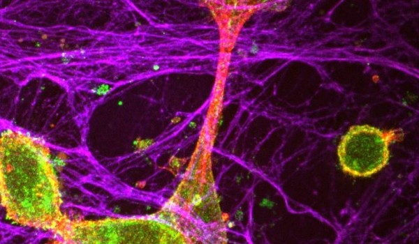
Triple-negative breast cancer cells are engaging extracelluar fibres (collagen, blue; actin, red) in a 3D matrix, which was grown in the lab using fibroblasts. The pro-invasive integrin aVb6 (green) is binding to and sensing the matrix, integrating information from the cell and its microenvironment. Cells can ‘grab hold of’ fibres and pull onto them to enable cells to migrate, and ultimately potentially invade into their surroundings…
Be aneuploid
Photographer: Federico Gulluni
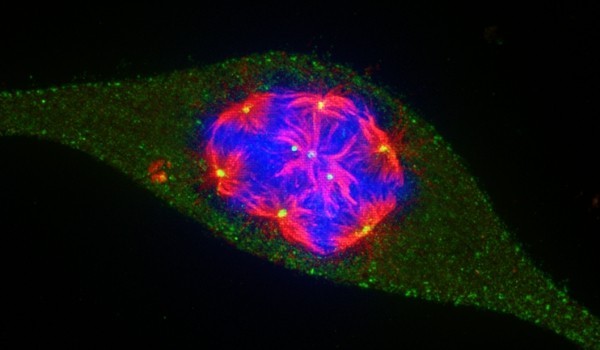
Immunofluorescence showing a cancer cell forming more than ten multiple spindles.
Blood vessels in a mouse brain
Photographer: Niraj Trivedi
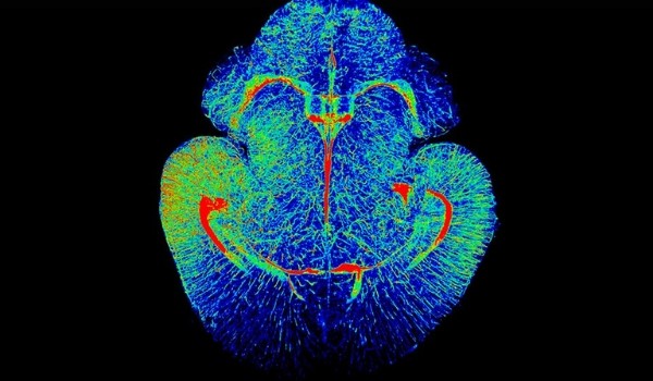
This is an image showing all the blood vessels in a mouse brain, 7 days after birth. The brain was isolated and treated using a cutting edge protocol called iDisco+, which makes the brain tissue completely see-through, giving it an almost a glass like appearance. This allows us to image deep into the tissue using immunocytochemistry combined with a LaVision Light Sheet Microscope to give this stunning image. The data collected was rendered using Arivis Vision4D software. The different colours reflect the intensity of the blood vessel staining (using a GLUT1 antibody). We also have the tools to visualize this data in virtual reality (VR) to see the nuances of the blood vessels in different regions and to make accurate measurements in VR.
The desperate struggle
Photographer: Anna Tosi
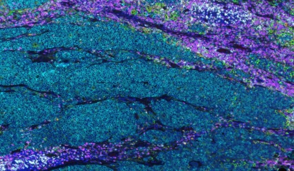
The image shows a lymph node metastasis of colon cancer in which tumor cells (in cyan) are invading the lymph node, while patient’s lymphocytes (in magenta and pink) try to restrain it.
Cancer Scientist is female and male
Photographer: Balkees Abderrahman
Women remain significantly underrepresented in science including cancer research, and their intellectual capacity or emotional fortitude are constantly questioned by others in such fields. This image honors the theme of equal-gender contribution in cancer research. Cancer science needs equally women and men to push the frontier of knowledge.
Striking Image S. haematobium
Photographer: Monica Botelho
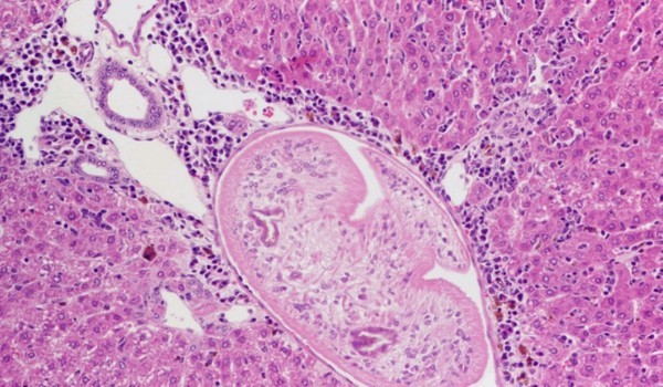
A Schistosoma haematobium worm trapped in the liver portal space of an infected hamster. Note the exuberant inflammatory infiltrate surrounding the vessel that contains the worm. This inflammatory infiltrate is characteristic of a granuloma, precursor of S. haematobium-associated cancer. Infection with S. haematobium is carcinogenic to humans (IARC Group 1 of carcinogenic risks to Humans).
Cancer can be somewhat FISHy
Photographer: Lisa Lee-Jones
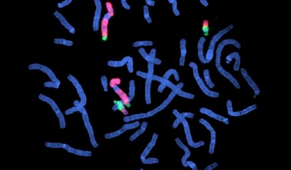
Reverse chromosome painted, flow sorted, der(9) and der(22) of the classical t(9;22)(q34;q11) rearrangement characteristic of Chronic Myeloid Leukaemia