Creating virtual reality maps of tumours: interview with Greg Hannon
In an exclusive interview, Greg Hannon from Cancer Research UK Cambridge Institute (UK), speaks to Oncology Central about his project to create virtual reality maps of tumors. The ground-breaking project has been selected by Cancer Research UK to receive £20 million over the next 6 years as part of its Grand Challenge awards; set up to revolutionize the prevention, diagnosis and treatment of cancer and help scientists tackle unanswered questions in cancer research.
Can you tell us about your career to date and your work at the CRUK Cambridge Institute?
I started off as a PhD student in Cleveland training in RNA biology and then moved to Cold Spring Harbor (NY, USA) 25 years ago, where I worked for 23 years on cell cycle control in cancer. That is where my interest in cancer started. As I transitioned to my own lab, also at Cold Spring Harbour, I maintained a dual interest and I was sucked in to the whole phenomenon of RNAi. I spent a lot of time working out mechanisms of RNAi, small, RNA biology, and microRNAs. I am still working on small RNAs that are a mechanism for defense of germline genomes.
We have always maintained our interest in cancer biology. A lot of the work that we did at Cold Spring Harbour was in collaboration with Scott Lowe, who is now at Memorial Sloan Kettering Cancer Center (NY, USA). There was also always a third current running in the lab which was that of technology development. I left CSHL approximately 2 years ago to move to Cambridge (UK). I also have a small lab at New York at the New York Genome Center (USA).
In some ways the move to the Cancer Research UK (CRUK) Cambridge Institute (UK) and having new colleagues has bought all of these interests together in to the Grand Challenge where there is a huge amount of technology and development [1]. By conducting this project we hope to learn a lot about tumor biology and at the same time come at this from the perspectives of measuring RNA copy number and examining RNA localisation in cells along with taking measurements of protein etc.
You are currently the principal investigator for the Grand Challenge: Finding a way of mapping tumors at the molecular and cellular level. Could you tell us about the aims of this project?
The project was born out of my exposure a number of years ago to work done by neuroscientists at Cold Spring Harbour particularly Pavel Osten who has helped to develop fluorescent serial-block face imaging two-photon microscope (STPT). He was looking in a great deal of detail at structures within the brain where certain reporters become active. This made me think that we could do the same thing in cancer biology to start to understand the 3D arrangement of cells in tumors.
At that point it was a hobby project and not the primary focus of the lab i.e. we would go and work with Pavel for a bit just trying to get our tissues fixed and sectioned and imaged appropriately.
Then with the move and with an almost a perfect coalescense of a set of colleagues here, we began to get much more serious about it and to think about taking what was really primarily an imaging project and turn it into something that was much bigger. From this project we hoped to understand not only the orientation of tumor cells vs something simple such as the vasculature but the arrangement of all the cell types in a tumor, their locations and then to add some deep molecular characterization.
This could allow us to identify each cell type cell, understand the activity of its signaling pathways and understand how different cell types behave when in contact within neighboring cell types.
We started working towards this and then the Grand Challenge questions were released and it seemed like mapping tumors in 3D was really a perfect fit. The project allowed us to think even bigger and to assemble a team of collaborators not just in Cambridge but to pull individuals from all over the world. Our project includes researchers from Vancouver, Dublin, Boston, Zurich and Lausanne plus a core group of five investigators back here in Cambridge. Overall, we were able to think with using a breadth of technology and doing analyzes on a scale that was unthinkable in a way prior to CRUK conceiving this funding mechanism.
What imaging methods will you use to develop the 3D tumor maps?
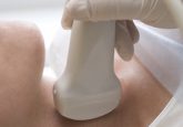
The first thing we do with each sample is to make a 3D reference model i.e. a model of the sample in its unperturbed state. There are two ways to achieve this. One is serial block face two-photon imaging. Serial two-photon tomography microscope is made by tissue vision, which scans alternating optical and physical sections. For example it will take a 100 micron optical section and then a vibratome will cut off a physical section.
It is then possible to re-image and stitch all of those images back together into a 3D whole. We would like to take the physical sections that are produced and characterize them molecularly. We have been working a lot with tissues to try to accomplish a way to section our tissues thinly enough and also to recover the sections that are made, which in the current configuration of the instrument would just float off into the buffer. Once we have the sections in hand we have three types of measurement modalities.
One is imaging mass cytometry, Bernd Bodenmiller (University of Zurich, Switzerland) is helping us push this angle. Imaging mass cytometry in essence CyTOF, which is a time of flight mass spectrometry, married to a FACS, conceptually at least. Cells are labelled with up to 50 different antibodies and shot one by one through a mass spectrometer, the abundance each the antibodies is then measured. What Bernd did was to convert this into an in situ method where a laser raster scans the tissue and you get measurements (up to approximately 50 different antibodies with one micron pixel sizes). This can be utilized to measure protein abundance and phosphorylation states etc.
A second measurement modality was developed by Xiaowei Zhuang (Harvard University; MA, USA) who is also a member of our Grand Challenge team. This is termed MERFISH (Multiplex error robust FISH) which is a way to do single molecule FISH and to read hundreds to thousands of different transcripts in a single cell. Again this is an in situ method so we do this on our tissue sections.
The third method that we are working towards is in situ sequencing. We have a collaborator, Ed Boyden, (MIT, Boston, USA) who has been working with George Church to adapt this in situ sequencing methods to his expansion microscopy methodology. To be able to expand the sample cells would solve a lot of problems potentially with the original in situ sequencing methodologies.
Those are our three current molecular characterization methods, after this we take information from each of those and assign it back to individual cells in the reference image. For that we have a great collaboration here with another member of our Grand Challenge team, Nicholas Walton (University of Cambridge, UK), who is an astronomer and of course what astronomers do is take images and recognize objects and assign parameters to those objects.
We are hoping that by bringing together all these individuals who have developed these cutting edge molecular characterisation methods, with some of the 3D imaging methods, with individuals who are very good at putting all of this information together that we will be able to create these 3D annotated maps.
We also need ways to interact with the maps; this led to our collaboration with our virtual reality group led by Owen Harris (Súil Design; Dublin, Republic of Ireland) who will integrate all of this information into a viewer. We have members of the team for example Carlos Caldas (Cancer Research UK Cambridge Institute, UK) and Sam Aparicio (University of British Columbia, Vancouver, Canada), who bring a detailed expertise in breast cancer which is the initial focus of our project and also a lot of really amazing technologies in their own right as well as lots of patient derived xenograft models, and single cell technologies that Sam Aparicio has developed. In our Grand Challenge team we also have Johanna Joyce (University of Lausanne, Switzerland) ,our tumor microenvironment expert who as we learn more and more in terms of our investigation will really help guide the questions that we are asking, particularly in mouse models regarding the interactions of the microenvironment with tumor cells and vice versa.
Another member of our team is Shankar Balasubramanian (Cancer Research UK Cancer Institute) who was one of the originators of the Illumina sequencing technology. Not only is he an avid biologist, but the chemistry expertise that he brings will be incredibly important. There are also a lot of technology problems to solve to make this all happen that cut across the imaging/chemistry space top. His expertise has already been very important in the initial phases of the project. Then there is Simon Tavare (CRUK Cambridge Institute) who is our computational biologist and statistician. He is interested in how the lineage of cells influences their behavior and he has also helped to develop many of the higher order measures and statistical methods that we will use to detect new 3D interactions. So these are all the important individuals and then there’s me and I mainly help put all this together
In the long term, how would you like to see this model utilized (in the clinic/in the lab)?
Our initial goal really is to have this as a research tool, to allow us to interrogate tumors in a way that was previously impossible and to obtain many more measurements than have ever been measured before. Now we also have other goals to expand their range and applicability of these models,we certainly see that this is potentially a very powerful educational tool for everyone from university students to training doctors and also very importantly patient education. We are developing multi-user environments where individuals can actually meet inside virtual space even if they are not physically in proximity and examine these models. We have two patient advocates who are leading this part of the project. We can imagine a doctor going in with a newly diagnosed patient, not necessarily with their own tumor but with a highly representative model of their subtype and explaining why they would be treated in a particular way. I can also see this in science museums etc for educating the general public. Our long term goal is to see this used in some way in the clinic. It probably wouldn’t be a full implementation as this would require tens of thousands of measurements about every cell. However, it could probably be informed by all of the work that we do to make a shortened list of measurements to give clinically actionable information. The fantasy would be that this becomes the new pathology which is both the visual structure of the tumor and comprehensive molecular classification characterization.
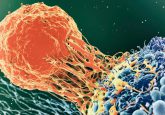
You have previously likened tumors to an ecosystem. Could you tell us more about this concept and why it is so important to view tumors in this way?
So we already know that tumors are heterogeneous. They are heterogeneous from patient to patient even with the same organ of origin. They are heterogeneous, within the tumor- different cells within tumors have different genetics, and more and more we are realising that different cells within tumors have different capabilities and different proficiencies. There is also a whole variety of different cell types that in many studies have been associated with differences in outcomes. Certain kinds of immune infiltrate give you a better or worse prognosis, what we are trying to do is to take that sort of high level observation and dig down to the underlying cause of this change. Is because this kind of immune cell in the tumor is interacting with tumor cells in this way? Or perhaps this kind of immune cell is being instructed by the tumor cell to act in a certain way? This system of tumor communication and competition is very much like an ecosystem which obviously is going to be perturbed by treatment. Currently we do not know a lot, certainly not in the 3D setting about how treatment influences the structure of the tumor. From this we hope to discern anything during that process that will enable us to make better predictions about how to apply therapy.
Could you tell us about the discoveries you have made in the field of cancer?
We have a long history of working in non-coding RNA biology. For example we have done a lot of work on microRNAs acting as oncogenes and working tumor suppressor networks. However, for the last 6 -7 years the majority of our thrust in the non-coding RNA space has been to try to understand the biology of the small RNAs in germ cells. This is probably not terribly cancer relevant but we have some upcoming projects that are highly cancer relevant in the long non-coding RNA space. Hopefully we will have a lot more to say about those soon.
In the cancer space we’ve done a number of different things. Early on in my career I was involved in linking regulators of the cell cycle to cancer as tumor suppressors. For example P16, P15, P21etc. As we went on we worked to develop tools that have been broadly used in the cancer community as shRNA etc. I would highlight our work on microRNA as oncogenes and a lot of work with with Scott Lowes lab at CSHL, trying to apply things that we would learn in the small RNA space particularly as tools in the cancer area. There were also some other developments worked on by us and at the same time by Richard Gibbs lab that had a big impact on the cancer space allowing individuals to conduct exome sequencing. More recently we have become very interested in tumor heterogeneity.
We to try to look at it in a functional way rather than in a retrospective way. Lots of researchers conduct experiments on tumor heterogeneity, initially single cell copy number analysis and single cell sequencing cataloging the diversity of genotypes in tumors. So what we’ve been trying to do is to catalogue the diversity in the phenotypes. To achieve this we have been building functional models of tumor heterogeneity and then probing the models to identify cells with very specific proficiencies to try to understand the molecular basis of phenotypic differences. An example, it was probably 2 years ago we published this, we created a model of breast tumor heterogeneity in mice. We identified a subset of cells that were very proficient at vascular mimicry. We traced this to a molecular basis and have been extending these observations both in terms of a mechanistic understanding of what drives vasculature mimicry and in terms of trying to uncover other subpopulations in that hetergeneous model that demonstrate very specific proficiencies. This is involved in a more recent study that is in consideration for publication about regulators of proficiency for colonization at secondary sites within the model.
What are you excited about working on over the next few years?
Well obviously I am excited about working on the Grand Challenge or else they probably wouldn’t have given me £20 million! I think actually that it is going to be a phenomenal adventure. It combines so many different interdisciplinary threads to try to do something in biology that has never been done. Although certainly our application initially is cancer, all of biology happens in a 3D context , development for example and this is something that I think would be very important to use the same kinds of tool kits that we will be building to understanding tumor biology. So what I’m really excited about is putting this all together and learning something about breast cancer but also building the foundation that we and lots of others can use to understand many other processes in biology.
Do you have any closing comments for our readers?
Stay tuned, it’s going to be interesting to see how this turns out.
References
[1] Cancer Research UK – Creating a virtual reality of tumours www.cancerresearchuk.org/funding-for-researchers/how-we-deliver-research/grand-challenge-award/funded-teams
Profile:
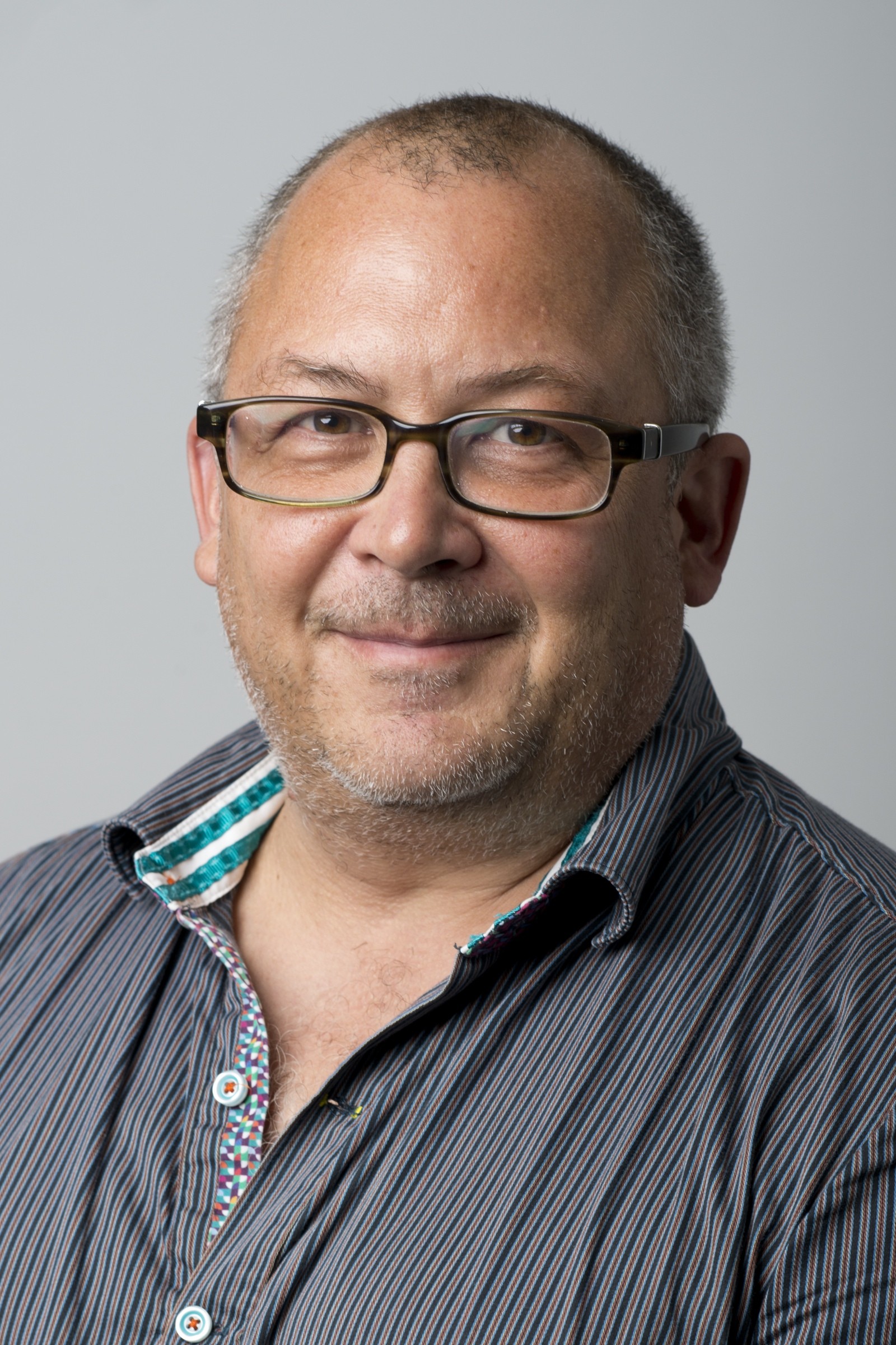
Professor Greg Hannon is internationally recognized for his contributions to small RNA biology, cancer biology and mammalian genomics. He has a long history in the discovery of cancer genes and has developed widely used tools and strategies for manipulation of gene expression in mammalian cells and animals, including generating genome-wide shRNA libraries that are available to the cancer community.