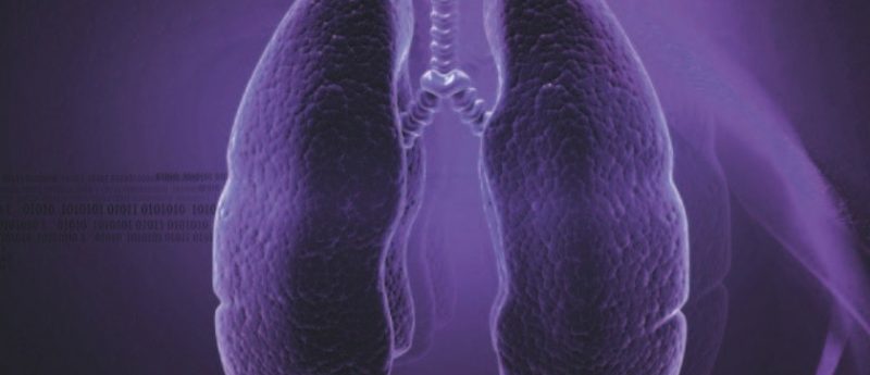
New technology developed to improve tumor detection in lung cancer
Researchers at Loyola University Chicago Stritch School of Medicine (IL, USA) have developed a new type of imaging that improves the detection of tumors during radiation therapy for early-stage lung cancer. John Roeske and colleagues reported the results of the study at the annual meeting of the American Society for Therapeutic Radiology and Oncology (14–17 September, San Francisco, CA, USA).
The new technology combines dual-energy imaging with fluoroscopy to visualize tumors during radiation therapy. There is no requirement for an x-ray, and visualization of the tumor is made easier by existing hardware that can remove visuals of bones.
“Dual-energy imaging has been used for decades by radiologists to detect lung tumors,” explained Roeske. “When combined with fluoroscopy, the hybrid dual-energy technology can enhance the visibility of tumors to improve treatment for patients.”
The researchers have a patent on the technology and predict that commercialization could mean cost reductions for hospitals.
“This technology does not require that hospitals replace their standard x-ray machines since the dual-energy images are created using a software approach,” Roeske commented. “The hybrid technique removes present obstacles making this a great benefit to clinicians and patients.”
Source: Loyola University Chicago Stritch School of Medicine press release